 positive
  request CD with images
TransfluorTM Image Sets
1. CompuCyte image set
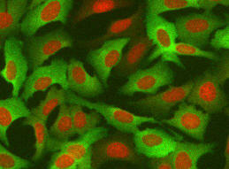
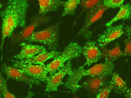
This image set has a portion of a 96-well plate containing 3 replica rows and 12 concentration points of isoproterenol. In each well four fields were acquired. The images are of U2OS cell line co-expressing beta2 adrenergic receptor (b2AR) and arrestin-GFP protein molecules. The receptor was modified-type that generates "vesicle-type" spots upon ligand stimulation. The plate was acquired on iCyte imaging cytometer with iCyte software version 2.5.1. Image file format is JPEG with one image for green channel and one image for crimson channel. Image size is 1000*768 pixels. File name structure: <well-number>_<field>_<channel>.JPG
negative  positive
  request CD with images
2. Roche image set
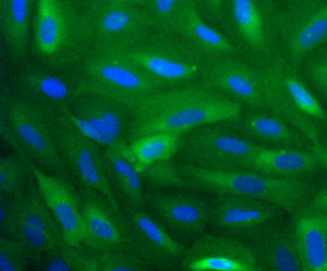
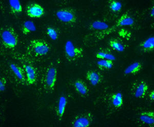
This image set is of Transfluor assay where an orphan GPCR is stably integrated into the b-arrestin GFP expressing U2OS cell line. After one hour incubation with a compound the cells were quenched with fixative (formaldehyde) and the plate was read on Cellomics ArrayScan HCS Reader using the GPCR Bioapplication. The images constitute one row of a 348 well plate. The dose curve consists of 11 dose points and one control. Each concentration is duplicated in adjacent wells. Each well has three fields. File format is 8-bit TIFF with one image for green channel and one image for blue channel. Image size is 512*512 pixels. File name structure: <prefix>_<row><column><field><channel>.TIF
negative  positive
  request CD with images